Thankfully there has been a revolution in the world of equine dentistry in the last 15-20 years such that a quick rasp ‘while you are here…’ with one or two battered old rasps, often no gag, and certainly no headtorch in sight, has become a thing of the past. The importance of regular, thorough dental examination and routine treatment in the maintenance of health for equines of all ages has been widely acknowledged. The development and improvement of treatment techniques and dental equipment has led to significant advances in the types of treatments available to help our equine friends.
Who can perform an equine dental?
The term ‘Dentist’ is not protected, which means that anyone can advertise themselves as an ‘Equine Dentist’ and attend patients, with no required qualifications or regulation. It is important to ensure that the professional attending your horse is suitably trained and qualified to ensure the safety and welfare of the horse1. Dental professionals should be qualified as an Equine Dental Technician (EDT) or a Veterinary surgeon. Registers of EDT’s and Veterinary surgeons with further certification in equine dentistry are available on the British Equine Veterinary Association (BEVA) website https://www.beva.org.uk/Guidance-and-Resources/Routine-Healthcare/dentistry.
How often are equine dental examinations needed?
Routine dental examinations are typically first carried out on young horses prior to them starting ridden training, at around 3 years old. It is generally advised to check these young horses twice yearly, as they are still maturing, with the cheek teeth not fully erupted until between 4-5 years. This enables us to detect and treat developmental issues such as retained deciduous teeth (‘caps’ – baby teeth which are shed as the adult teeth erupt) and wolf teeth, which may or may not require removal depending on their location and presentation. Young horse teeth also develop sharp edges quickly and it is important that these are addressed to maintain their comfort when wearing bits and bridles. Horse teeth will grow to their full length by around 5 years, with most of the tooth below the gum at this point. The teeth then continually erupt throughout the horses’ lifetime, until the tooth has such little reserve left below the gum that it tooth becomes loose and may fall out. Mature horses without any ongoing dental problems may be managed satisfactorily with once yearly examinations. Older horses or those with dental disease are likely to need more frequent examinations and treatment.
What does a dental examination entail?
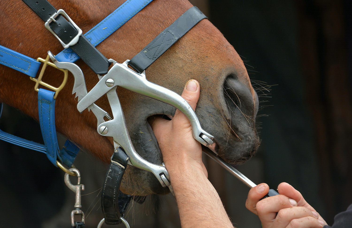
A thorough dental examination will include the use of a speculum (a ‘gag’), with a light-source and either a mirror or even better an oroscope (camera) to visualise the whole mouth. The horse may rest their head on a head-stand to help improve visibility. It is advised that horses are sedated to facilitate this full examination for safety (of both horse and handlers) and to minimise movement for the time required, so that the examination is not rushed or incomplete.
What signs of dental disease can we look out for?
Potential signs of dental disease which should be noted and investigated include:
- ‘Quidding’ – dropping balls of partially chewed food
- Smelly nasal discharge – usually from one nostril
- Weight-loss or failure to gain weight
- Contact issues or one-sidedness when ridden
- Behavioural issues – including irritability when handled or unpredictable or violent behaviour under saddle.
It is important to note that often dental disease is present with no apparent signs reported – horses are very good at hiding pain!
What can go wrong in a horse’s mouth?
There are several frequently encountered issues which can cause pain and discomfort for the horse2.
The most common is the sharp enamel edges which form primarily on the outer edges of the upper teeth and the inner edges of the lower teeth. These can cause ulcers on the cheek and tongue, and often the horse will be less keen to take the contact with the bit when ridden. It is these edges which are rasped smooth every 6-12 months.
Other common abnormalities which may be detected in the mouth include caries (cavities) 3, diastema (gaps between teeth which trap food and cause gum disease – these are the most painful abnormalities we see)4, fractured teeth, and Equine Odontoclastic Tooth Resorption and Hypercementosis (EOTRH)5. EOTRH is a degenerative condition usually affecting the incisors (front teeth) of older horses. It causes tooth root reabsorption and is often associated with gum swelling and pain. These teeth become loose and are a cause of discomfort. The only treatment possible for EOTRH at present is extraction of the affected teeth.
Recent advances in investigation and treatment of equine dental disease
The detection of dental disease and the accuracy of diagnosis has been hugely improved by the ability to carry out head CT scans in the conscious horse. These scans are invaluable for detecting tooth-root infections which can be difficult to diagnose with examination and X-Rays alone6.
Dental treatment options for horses have also developed in recent years, extraction is not the only treatment we can offer any more!2. Dental specialists can offer fillings to treat cavities3, perform root canal treatment to treat damaged or infected pulp cavities, and diastema widening. For those teeth which do unfortunately need extraction, almost all can be removed without the requirement for a general anaesthetic.
References:
- BEVA website current regulations on Equine Dental procedures:
- Equine dentistry – common problems and solutions
Neaera Fletcher Veterinary Practice Today p38-39 Vol. 8, Issue 2, 2020
- Equine dental caries and restoration Jennifer E. Rawlinson Equine Veterinary Education p621-626, Vol.35 Issue 12, December 2023
- Is flushing and packing adequate for diastema treatment? N. Townsend Equine Veterinary Education p385-386, Vol.27, Issue 7 July 2015
- Radiological prevalence of equine odontoclastic tooth resorption and hypercementosis S.Rehrl, W.Schröder, C. Müller, C. Staszyk, C. Lischer Equine Veterinary Journal p481-487, Vol. 50, Issue 4, July 2018
- Radiographic, computed tomographic, gross pathological and histological findings with suspected apical infection in 32 equine maxillary cheek teeth (2012–2015)
T. Liuti, S. Smith, P. M. Dixon Equine Veterinary Journal p41-47, Vol.50, issue 1, January 2018