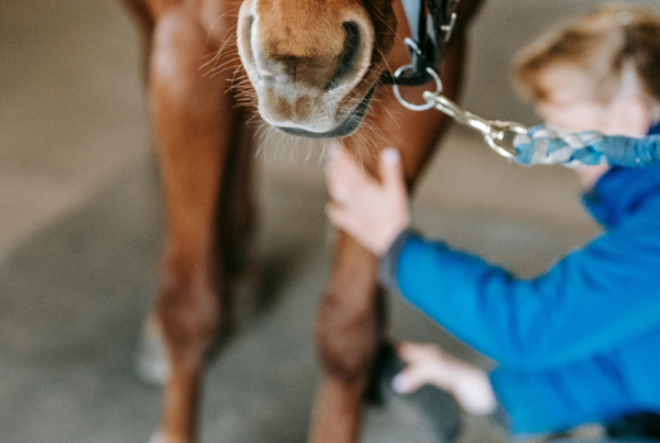
Routine dental examinations are one of the most important healthcare gifts we can give to our horses. Horses are extremely good at hiding any signs of dental disease and just because your horse is eating well or managing to maintain (or even exceed!) an ideal weight, does not rule out the presence of disease. It has been reported that up to 90% of horses will have some degree of dental problems, which can range from a few sharp enamel points to advanced fractures.
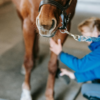
In recent times, advances in equipment means that we can detect signs of disease at an early stage, which means that treatment is often easier and potentially more successful. In the practice, we aim to examine horses’ teeth at least once a year, more frequently if the horse is older or has a history of dental problems. We will always use a full mouth speculum and a headtorch and mirror. While this may look like a lot of kit for “a quick check”, in fact it allows us to examine the mouth thoroughly, and means we can catch issues before they become major problems.
Do horses get dental decay?
Yes they do! Historically we used to think that, like dogs and cats but unlike humans, dental decay was rare in horses. However, newer research over the last few years has shown that actually horses suffer from not just one, but two different types of dental decay or caries:
Peripheral caries
This is a type of decay that is found on the sides of the teeth. It starts as erosion into the outer surfaces of the tooth and may be so mild that it just appears as a discolouration and pitting of the surface of the tooth.
Infundibular caries
This is decay that occurs in a particular part of the tooth, the infundibulum. This is a deep, enamel-filled cup on the chewing surface of the upper cheek teeth, and incisors, although incisors are rarely affected by caries.
What causes peripheral caries (PC)?
Peripheral caries are very common in horses, a recent large-scale study found that they were present in almost 52% of horses. Mares tend to be affected more frequently than geldings or stallions.
Peripheral caries (PC) are thought to be caused by bacterial erosion of the surface cementum of the tooth. We see PC more frequently in the back teeth, and a possible reason for this is that the salivary duct is located near to the front of the mouth, so teeth at the front are covered with acid-neutralising saliva to a greater extent than the back teeth.
Interestingly, horses with PC are more likely to have other dental problems such as gaps or diastema between teeth. It is possible that dental problems like sharp enamel points allow food to get trapped on the sides of the teeth, and their tongues are not very effective at removing food stuck at the back, which in turn allows for bacteria to grow, develop and cause erosions of the tooth. If left untreated, decay can develop and destroy the tooth.
How can peripheral caries be treated?
With the above factors in mind, treatment of PC involves reducing the acid load on the teeth and ensuring that food can move freely in the mouth, reducing the risk of food sitting and rotting on the surface of the teeth.
One way of helping this is to carry out a thorough dental rasping and treatment of underlying problems, to maximise the chewing potential of the teeth and to reduce the risk of pockets of food developing.
In addition to dental treatment, we can also examine the diet to see if there is an excess of acid-rich food. Dietary management may help to delay the spread of PC. Checking the water source may also be advisable, to ensure that the pH is as close to neutral as possible.
If your horse will tolerate it, rinsing the mouth with fresh water from a hosepipe several times a day will help to remove any stagnant food.
Fluoride gels or paints are currently under investigation and may form part of treatment in the future.
The positive thing to bear in mind with PC is that they can improve over time. The horse’s teeth erupt continuously during life and if management changes are implemented, along with regular dental care, it is possible to delay the progression or even reverse the severity of any lesions. We usually aim to follow up cases regularly.
What about infundibular caries (IC)?
Infundibular caries are also very common in horses, and the incidence tends to increase with age. As mentioned previously, these are found on the chewing surface of the upper or maxillary teeth. These are diagnosed visually, with the use of mirrors or a specialised camera, called an oroscope. The vet may also use a dental pick to determine the depth of the cavity. In advanced cases, more specialised imaging such as radiography or CT may be necessary to determine the significance of the decay. In the early stages, food packing is found in the infundibular cups, but as disease develops, the internal structure of the tooth may be weakened with the end result of severe decay being fracture and loss of the tooth.
How can infundibular caries be treated?
Early lesions may be treated by monitoring alone. Our vets will record and document any findings so that we can check to see if decay is progressing. Early caries may stay static for years, so advanced treatment is only recommended if the decay is worsening or of a high grade. In advanced cases, treatment by filling is used. This procedure is quite specialised and may necessitate referral and further imaging such as CT or X-ray. However, success rates in appropriate cases are very high.
In the first instance, the vets will make sure that they document any findings, using standard grading systems so that we can monitor your horse over time. In addition to this, regular, careful dental treatment to remove any underlying problems and management changes, will all help to delay the progression of the decay.
If you have any questions, or would like to book in a dental exam for your horse, please contact us where we would be delighted to help you.